| Cochlear
potentials |
| There
are 2 types of cochlear potentials : individual unit potentials recorded
directly from a sensory cell or nerve cell, and compound potentials, recorded
at a distance, reflecting the activity of several cells. |
Single unit potentials |
 |
Single
unit potentials recorded from the sensory cells are also known as receptor
potentials. |
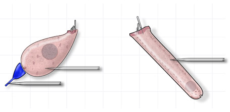
These
potentials are recorded via a micro-electrode placed directly into the
cell (intracellular recordings).Single unit potentials of the primary
auditory neurones can be recorded either at the level of the dendrites
(as shown), the cell bodies in the spiral ganglion, or at the level of
the auditory nerve fibres.
|
Hair cell
receptor potentials |
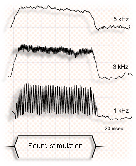 |
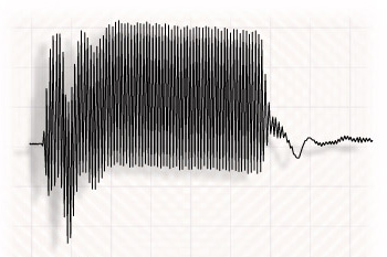
The
electrical response of the hair cells to an acoustic stimulus (shown
at the bottom of the figure) is made up of two components : a continuous
component (CC), duplicating the acoustic stimulus envelope, and
an alternating component (AC), superimposed on the CC, corresponding
to the pure sound frequency. These continuous and alternating components
depend on the frequency of sound stimulation. The amplitude of the
CC grows with the frequency, whereas the CA is more important for
low frequencies. The CA is more important in the outer hair cells
than the inner hair cells, and the CC is more dominant in the inner
hair cells (ref. Russell et al.). |
|
Auditory nerve
single unit potentials |
 |
|
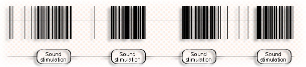
A
sound stimulation (click) leads to an increase inthe numbre of single
unit potentials in the individual fibres of the auditory nerve. This
increase in the rate of discharge, synchronised with the sound stimulation,
is called an evoked potential. The presence of action potentials during
periods of silence is a reflection of the basal activity of the nerve
fibre, also known as its spontaneous activity.
|
Composite
cochlear potentials |
|
|
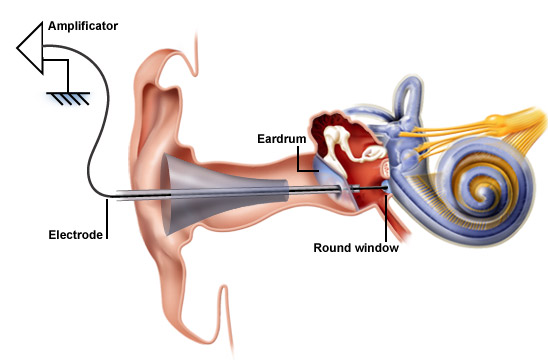
| Electrocochleography
(ECochG) is the name given to the recording of cochlear potentials.
Under local anaesthetic, a thin needle electrode is placed through
the tympanic membrane onto the promontory, near the round window
niche.(ref. Aran et al.) |
 |
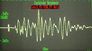
The gross
cochlear response complex is recorded in response to a sound
stimulus (i.e. it is an evoked response), and it is made up of
various components. The use of appropriate filters allows the
identification of these components, which in turn correspond to
the various anatomical structures implicated in the mechanotransduction
of sound and in the transmission of auditory information. |
Compound action potential (CAP) |
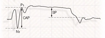 |
The compound
action potential (CAP) is the result of synchronous activity
of the auditory nerve fibres. The CAP amplitude is measured between
N1 and P1. The summating potential (SP) reflects the continous
component of the sensory hair cells, mainly the IHCs. |
Microphonic potential |
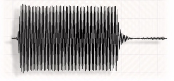 |
The cochlear
microphonic, which closely resembles the sound stimulus, is
a reflection of the alternating component, which mainly originates
from the OHCs.. |
|