| |
|
| Overview / Physics / Fluids / Stria | |
| Drawings: S. Blatrix; Pictures: M. Lavigne-Rebillard, M. Lenoir | |
| |
|
| Overview / Physics / Fluids / Stria | |
| Drawings: S. Blatrix; Pictures: M. Lavigne-Rebillard, M. Lenoir | |
|
|||||
 |
 Plan of cross section Plan of cross section |
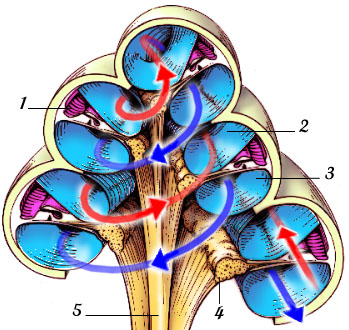
| This mid-modiolar section shows the coiling of the cochlear duct (1) which contains endolymph, and the scala vestibuli (2) and scala tympani (3) which contain perilymph. The red arrow is from the oval window, the blue arrow points to the round window. Within the modiolus, the spiral ganglion (4) and auditory nerve fibres (5) are seen. For details, see the single turn cross section below. |
|
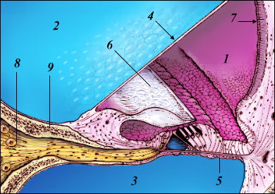 |
| The cochlear duct (1) is isolated from the scala vestibuli (2) and scala tympani (3) by Reissner's (4) and basilar (5) membranes respectively. The organ of Corti is covered by the tectorial membrane (6) floating in the endolymph. The stria vascularis (7) and the fibres (8) going to the spiral ganglion through the bony spiral lamina (9) are also shown. |
| View
of a rat cochlear spiral (Scanning electron microscopy) |
|
 |
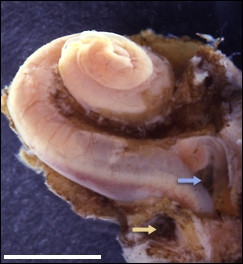
In the rat, the basilar membrane develops a 3 turn spiral of a total length of 22 mm (compare with the coiling in human). Only the extreme basal part (the hook) is not seen. scale bar: 2 mm Note that the bony capsule, stria and tectorial membrane have been removed |
| M. Lenoir |
|
 |
For
permission to non-commercial use of any element of this site, please contact us All rights reserved © 1999 - 2007 The authors Intellectual property law 85-660 (07/03/1985) |
|