|
|
|
| Overview / Function / Development | |
| Drawings: S. Blatrix; Pictures: M. Lavigne-Rebillard, M. Lenoir, R. Pujol | |
|
|
|
| Overview / Function / Development | |
| Drawings: S. Blatrix; Pictures: M. Lavigne-Rebillard, M. Lenoir, R. Pujol | |
| In humans, the organ of Corti begins its differentiation at 9 weeks of gestation (wg). The following set of Nomarski and SEM pictures illustrates the main stages of its development (ref. b24). A timing correspondence between humans and different mammals is given in table I. |
Stage 3 :
Organ of Corti just prior to the onset of function
(14-15 wg)
|
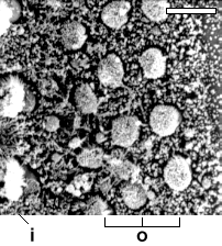
SEM surface of
a 14 wg human fetus cochlea. Stereocilia of both IHCs (i) and OHCs (o) have grown and the
tufts have lost the round-shaped
pattern. scale bar : 10 µm |
 M.Lavigne-Rebillard |
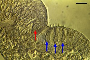
Nomarski section from a 14 wg human fetus. IHC (red arrow) and OHCs (blue arrows) are clearly distinguishable on both sides of the differentiating pillar cells. scale bar: 20 µm |
Similar developmental stage in a 2 postnatal day (pnd) old mouse. Note
the inner sulcus filled with the thick Kölliker's organ
(asterisk) which plays a major role in forming the tectorial
membrane by secretion (white arrow). scale bar: 20 µm |
 M.Lenoir |
 R.Pujol R.Pujol |
In all mammals studied, this stage corresponds to the onset of cochlear function (see table I). At this stage (here a pnd 8 mouse), the tunnel of Corti is open, Nuel's spaces are forming beside the OHCs (blue arrows), and Kölliker's organ (asterisk) is regressing, freeing the inner spiral sulcus and the tectorial membrane. In this section, the IHC is not fully visible. |
 M.Lavigne-Rebillard |
 M. Lavigne-Rebillard |
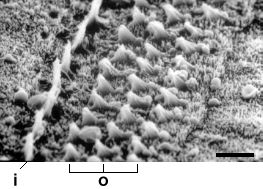
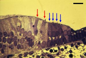
| Supernumerary hair cells are
frequently and temporarily encountered during the development of the human
organ of Corti, between stages 3 and 4 ( ref
c4). As shown here (14 wg, left; 20 wg, right) 2 rows of IHCs (red
arrows) and 4 or 5 rows of OHCs (blue arrows) are often seen, even in
the basal turn. scale bar: 20 µm |
|
Stage 5 :
End of morphological differentiation of the organ of Corti
(30
weeks' gestation in the human)
|
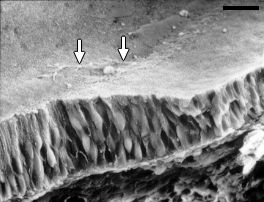
SEM picture (rat organ of Corti). At the end of its development (30 weeks of gestation) the human organ of Corti looks like this one (taken from a one month-old rat).Note an almost complete disappearance of microvilli on the surface of the support cells, especially those of the pillar cells. scale bar: 15 µm |
 R.Pujol |
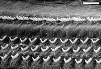
Mature organ of
Corti from a guinea-pig (Nomarski). The main differences between stages 4 and 5 concern the final stages of differentiation of the OHCs and surrounding structures. scale bar : 10 µm |
| This table gives
comparative timings of stages 2 (onset of hair cell morphological differentiation),
stage 4 (onset of cochlear potentials), and stage 5 (end of maturation)
in different mammals (ref b21). wg = weeks of gestation (humans) ; ed = embryonic days ; pnd = postnatal days |
|||
|
|
|
|
|
|
Human Rat, Mouse Cat Guinea-pig Gerbil |
10 wg 16-17 ed ? (ed) 34 ed ? (ed) |
18 wg 8-10 pnd 3 pnd 54 ed 12 pnd |
30 wg 16-20 pnd 20 pnd 6 pnd 20 pnd |
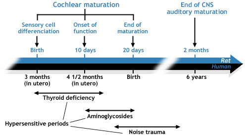 |
| This table recalls
stages 2,4, and 5 (see above) of cochlear maturation and highlights
the late development of the auditory brain. Actually, to
mature properly, the brain requires the cochlea to be
fully mature and functional. The hypersensitive periods to three major deleterious factors of cochlear development are indicated. During these periods the cochlea is much more susceptible than in adulthood (cf. ref.b11, b2). |
 |
For
permission to non-commercial use of any element of this site, please contact us All rights reserved © 1999 - 2007 The authors Intellectual property law 85-660 (07/03/1985) |
|
 .
.