Organ of Corti
Overview / Function / Development
 Drawings: S. Blatrix; Pictures: M. Lenoir, R. Pujol


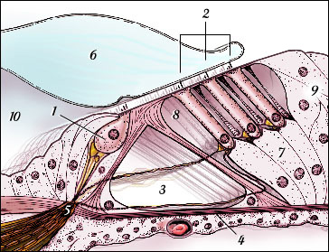
The organ of Corti is named after one of the first anatomists to give a detailed description (ref. a11) of the neuro-sensory cochlea. Seated on the basilar membrane, it is composed of the sensory cells, called hair cells, the neurons, and several types of support cells.

Schematic drawing of the organ of Corti


1-Inner hair cell
2-Outer hair cells
3-Tunnel of Corti
4-Basilar membrane
5-Habenula perforata
6-Tectorial membrane
7-Deiters' cells
8-Space of Nuel
9-Hensen's cells
10-Inner spiral sulcus

In this schematic drawing from a transverse section of the basal turn of a mammalian cochlea, the two types of sensory cells: inner (IHC: 1) and outer hair cells (OHCs: 2) are seen on both sides of the tunnel of Corti (3) which is limited by the 2 pillars. The tectorial membrane (6), floating in endolymph, covers the hair cell, embedding the tips of the tallest OHC stereocilia. The IHC is surrounded by support cells while the OHCs are firmly "seated" on Deiters' cells (7), their lateral membrane being in direct contact with the corticolymph (almost identical to the perilymph) which fills in the tunnel of Corti (3) and the spaces of Nuel (8).The cuticular plate of hair cells, together with the head of pillars, the phalangeal processes of Deiters' cells and the apical membrane of other support cells, e.g. Hensen's cells (9), form the reticular lamina (5) sealing the endolymphatic compartment. Piercing the basilar membrane (4) at the habenula perforata (5), nerve fibres reach or leave the organ of Corti .

SEM of guinea pig organ of Corti (mid-basal turn)
 
 M. Lenoir

Both the surface of hair cells and the inside of the organ of Corti (at site of fracture) are seen. Lateral to the OHCs, remnants of the marginal net of the tectorial membrane (which has been removed) are visible.

Blue arrows point to OHCs bodies, the asterisk indicates the tunnel of Corti where nerve fibres are crossing - the biggest (red arrow) are medial efferent fibres, the thinnest (green arrow) running on the floor of the tunnel are spiral afferent fibres

scale bar: 20 µm.


Transverse sections of the organ of Corti with Nomarski optics

 
 M. Lenoir

Mid basal (bottom) and third (top) turns from a guinea pig cochlea are shown. Compare with SEM pictures from the base and apex. 

See schematic drawing for legends.

The main morphological differences between the two turns concern the length of OHCs, regularly increasing from base to apex, as does the width of the angle formed by the reticular lamina and the basilar membrane.

Note also a hypertrophy of Hensen's cells with lipid inclusions (black spots) in the apical picture.

scale bar: 20 µm

 
R Pujol

For permission to non-commercial use of any element of this site,
please contact us
All rights reserved © 1999 - 2007 The authors
Intellectual property law 85-660 (07/03/1985)