| Overview / Synapses / Physiology | |
| Drawings: S. Blatrix; Pictures: R. Pujol | |
| Schematic drawing of an inner hair cell (IHC) | |
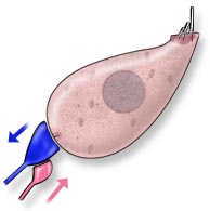 |
The IHC and its innervation are schematically represented here on the left. Note, unlike in OHCs, the medial location of the nucleus, and a "normal" lateral plasma membrane. Only a single synaptic complex is represented: i.e. a radial afferent bouton (blue) and a lateral efferent ending (pink). On average, one IHC is innervated by ten of these complexes. |
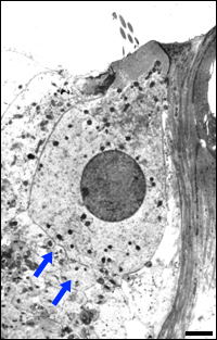
| Transmission electron micrograph of an IHC from a guinea pig cochlea | |
 R.Pujol |
Note the medially located nucleus and the randomly dispersed mitochondria within the IHC. Afferent nerve profiles (arrows) are seen at the base. On average, an IHC is connected by ten boutons from afferent dendrites. However, this number increases in the region of best frequencies. For instance, in the "fovea" of a bat cochlea (the portion of the cochlea coding for and around the echo frequency) up to 50 boutons per IHC can be observed. scale bar: 5 µm |
