| Overview / Coupling / Membrane / Synapses / Active mechanism / Oto-acoustic emissions | |
| Drawings: S. Blatrix; Pictures: M. Lenoir, R. Pujol |
| Overview / Coupling / Membrane / Synapses / Active mechanism / Oto-acoustic emissions | |
| Drawings: S. Blatrix; Pictures: M. Lenoir, R. Pujol |
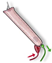 |
Below an inferiorly located nucleus the synaptic compartment contains mitochondria. An afferent bouton (green) from a type II ganglion neuron and a medial efferent ending (red) are represented at the synaptic pole of the OHC. |
| OHC
length variation according to frequency (adapted from ref. a25) |
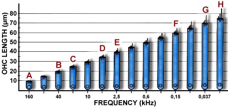 |
| Schematic drawing representing OHCs from different mammalian species and different cochlear turns. While OHC diameter keeps a constant value (7 µm), their length regularly varies according to frequency. In the human cochlea, a 25 µm basal OHC (C) is found at a place which codes for 20 kHz; conversely a 70 µm OHC (G) is found apically at the site coding for a very low frequency (< 100 Hz). A = shortest OHC in basal turn of a bat cochlea (at a place coding for 160 kHz), B = basal OHC from a cat cochlea (at a place coding for 40 kHz), D = OHC from second turn of a guinea pig cochlea (at a place coding for 5 kHz), E = OHC from start of third turn of a guinea pig cochlea (at a place coding for 2.5 kHz), F = OHC from end of third turn of a guinea pig cochlea (at a place coding for 150 Hz), H = apical OHC from a mole rat cochlea (at a place coding for 15 Hz). |
| OHCs
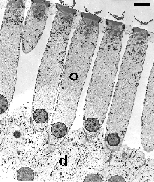
in longitudinal section: second row of apical turn in the rat cochlea (transmission electron microscopy) |
|
|
Very regular cylindrical-shaped OHCs (o) are seated on Deiters' cells (d). Their lateral membranes are separated from each other by the spaces of Nuel. The plane of section shows the "W" patterning of the 3 rows of stereocilia: this is better seen in the enlargement below. scale bar: 5 µm |
|
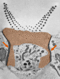
Enlargement of the cuticular plate (enhanced in brown) of an OHC from the above picture. The plane of section allows a precise view of the "W" pattern and a count of 3 rows of stereocilia. Also, the tight coupling with adjacent Deiters' cells is clearly visible (red arrows); this seals the junction between endolymphatic and perilymphatic (spaces of Nuel) compartments. |
| Cuticular plate and stereocilia from of a rat basal OHC (scanning electron microscopy) |
|
 M. Lenoir |
3 rows of graded length stereocilia are arranged in a "w" shape. The cuticular plate (internal to the stereocilia) is almost devoid of microvilli, contrasting with the surface of the surrounding support (Deiters') cells. scale bar: 1 µm See also the patterning and links of stereocilia in the mechano-transduction page. |
 |
For
permission to non-commercial use of any element of this site, please contact us All rights reserved © 1999 - 2007 The authors Intellectual property law 85-660 (07/03/1985) |
|
 R.Pujol