| |
|
| Overview / Coupling / Membrane / Synapses / Active mechanism / Oto-acoustic emissions | |
| Drawings: S. Blatrix; Picture: R. Pujol | |
| Electromotile properties of OHCs are due to the their very specific lateral membrane and sub-plasma membrane cytoskeleton. |
| |
|
| Overview / Coupling / Membrane / Synapses / Active mechanism / Oto-acoustic emissions | |
| Drawings: S. Blatrix; Picture: R. Pujol | |
| Electromotile properties of OHCs are due to the their very specific lateral membrane and sub-plasma membrane cytoskeleton. |
 R. Pujol |
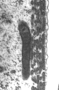
Endoplasmic reticulum cisternae are aligned along the OHC lateral membrane. Their numbers vary from 1 single row at the base to 5 to 6 rows (as in this picture from the third turn of a guinea pig cochlea) at the apex. |
|
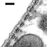 |
Same cochlea, basal turn. Between the
plasma membrane and the single row of cisternae, the lattice
of cytoskeleton can be distinguished: especially the transverse pillars. R. Pujol |
|
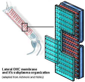
| Schematic drawing of the OHC lateral membrane and subplasma complex (adapted from refs. d3, d4) | |
Schematic arrangement of the OHC plasma membrane (green,) the subplasma membrane cytoskeleton (red), and the laminated cistern (blue). See electronic zoom for details. |
|
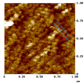
| Atomic force microscopy of the OHC lateral membrane (outer surface) (from ref. d7) | |
This scan on 1 µm² of an OHC membrane allows to distinguish a very specific arrangement of globular particles aligned in two perpendicular directions. Most of these are hollowed and could well be transmembrane proteins such as the the prestin motor? |
 R. Pujol |
 |
For
permission to non-commercial use of any element of this site, please contact us All rights reserved © 1999 - 2007 The authors Intellectual property law 85-660 (07/03/1985) |
|