Overview / Spiral Ganglion / Neurotransmitters |
|
| Drawings: S. Blatrix | |
Overview / Spiral Ganglion / Neurotransmitters |
|
| Drawings: S. Blatrix | |
|
|||||
| Innervation of inner (1) and outer hair cells (2) |
 |
| The radial afferents (blue) and the lateral efferents (pink) innervate the inner hair cells; the spiral afferents (green) and the medial efferents (red) innervate the outer hair cells. |
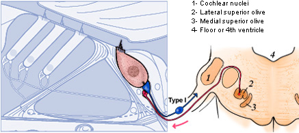
| Inner hair cell (IHC) innervation |
 |
|
The IHC is synaptically connected to all type I spiral ganglion neurons (refs. a1, c5) forming the radial afferent system (blue) going to the cochlear nuclei (CN). The lateral efferent system (pink) arising from small neurons in the ipsilateral lateral superior olivary complex (LSO) brings a feedback control to the IHC/type I afferent synapse (ref. c2, c3). |
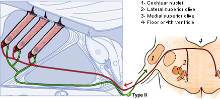
| Outer hair cell (OHC) innervation |
 |
|
The OHC synapses with a few (at least in basal and mid-portions of the cochlea) small endings from type II spiral ganglion neurons (ref. c1), forming the spiral afferent system (green). In turn, large neurons of the medial efferent system (red), from both sides of the medial superior olivary complex (MSO), form axo-somatic synapses with the OHC (ref.a2, a3). To better visualise the nerves within the organ of Corti, Deiters' cells have been removed. |
 |
Type I spiral ganglion neurons (95% of the ganglion neurons) have a single ending radially connected to IHCs. Type II small, unmyelinated neurons spiral basally after entering the organ of Corti and branch to connect about ten OHCs, generally in the same row. |

|
For permission to non-commercial use of any element
of this site, please contact us All rights reserved © 1999 - 2007 The authors Intellectual property law 85-660 (07/03/1985) |
|