Ear
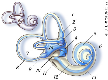
Overview / External ear / Middle ear / Inner ear 
Drawings: S. Blatrix


Vestibule and cochlea: two sensory organs of the inner ear

Two sensory organs are located in the inner ear. The vestibule is the organ of equilibrium and the cochlea the organ of hearing. They share a common embryonic origin (otic vesicle), plus different morphological or physiological properties such as endolymph, hair cells and mechano-transduction.
The schematic drawing below represents the osseous (top left) and membranous (seen by transparency in the main drawing) labyrinths.

 
1. Anterior semicircular canal
2. Ampulla (superior canal)
3. Ampulla (lateral canal)
4. Sacculus
5. Cochlear duct
6. Helicotrema
7. Lateral (horizontal) canal
8. Posterior canal
9. Ampulla (posterior canal)
10. Oval window
11. Round window
12. Vestibular duct (scala vestibuli)
13. Tympanic duct (scala tympani)
14. Utricule

 In situ schematic drawing of the human inner ear

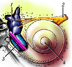
The bone has been removed to visualise the vestibule (1), the VIIIth nerve (2) , and the basal portion of the cochlear duct (3), housing the organ of Corti. The rest of the cochlea (4) is covered by the bony capsule. The VIIIth nerve is formed by the vestibular and cochlear nerves which merge before entering the brain.


Frequency distribution along the human cochlea basilar membrane: passive tonotopy
 

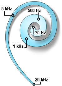
From base (20 kHz) to apex (20 Hz), some characteristic frequencies are indicated.
Note the progressive enlargement of the basilar membrane.

See also the animated drawings above.


  For permission to non-commercial use of any element of this site,
please contact us

All rights reserved © 1999 - 2007 The authors
Intellectual property law 85-660 (07/03/1985)
Début de page