| |
|
| Overview / External ear / Middle ear / Inner Ear | |
| Drawings: S. Blatrix; Pictures: M. Mondain | |
| |
|
| Overview / External ear / Middle ear / Inner Ear | |
| Drawings: S. Blatrix; Pictures: M. Mondain | |
|
|||||
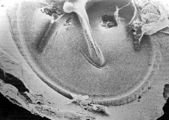 M. Mondain |
SEM picture of a guinea pig eardrum. The surface of the tympanic membrane is observed from the middle ear and the attached arm of the malleus is seen in the centre. |
| Ossicular reflex |
 |
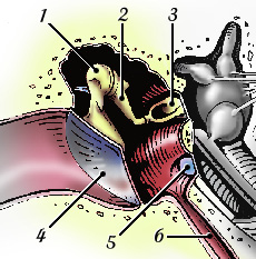
- (1) Malleus ; |
This
internal view of the middle ear cavity allows understanding of how the
ossicular reflex may reduce the transfer function of the ossicular chain.
This reflex protects the cochlea against loud sounds, but this type of
protection is quite limited:
Another function of the ossicular reflex is to attenuate perception of one's own voice: this is especially important for singers. |
|
Function of the middle ear
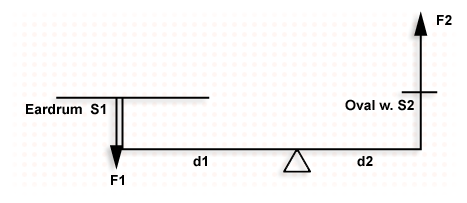
The middle ear (ME) transmits acoustic energy from the tympanic membrane (TM) to the inner ear, by allowing adjustment of the difference in impedance between an air environment and a fluid environment. If the sound pressure waves in the air were applied directly to the inner ear fluid, 99.9% of the acoustic energy would be lost because of their reflection at the air /fluid interface (equating to a loss of approx 30dB). |
|
The middle ear acts as a pressure amplifier: in this way it is able to “capture” the available acoustic energy in the air, and augment the amplitude of the mechanico-acoustic stimuli in the inner ear. Because of the relationship between the surface areas of the TM (area S1 = 0.6 cm2) and the stapes footplate (area S2 = 0.03 cm2), and because of the interaction of the ME levers (the axis of the ossicular chain passes very close to the incudomalleolar joint, but the two arms of this lever are of unequal length, d1/d2 = ~ 1.3), the pressure amplification is theoretically in the order of x26 (approx 28 dB) |
|
|
Beware, however! This calculation must be used with caution, because, due to its mechanical characteristics, the behaviour and the efficiency of the ME varies greatly with varying frequency of sound (f). In effect, ME function (similar to any type of mechanical system) depends on the friction (R) of the ossicular joints, the mass of the drum/ ossicular chain, and the rigidity (K) of the various membranes, ligaments, air volume and so on (see below).
|
||||
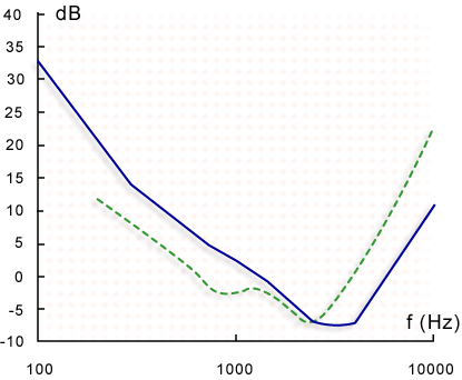
The mechanisms described above become obvious when one studies the transference function of the ME, ie the complex interaction between amplitude and phase that exists between the acoustic pressure at the entrance to the inner ear (Pv: pressure within the perilymph at the base of the scala vestibuli) and the pressure at the TM (Pt): Pv/Pt. In humans, the maximum sound amplification possible is 20dB, and this varies greatly with frequency, for example there is a maximum of 13dB amplification at 200 Hz, 20 dB at 1000 Hz, 12 dB at 8000 Hz.
NB In the range of audible frequencies, the impedance at the entrance of the cochlea (Zc) is purely resistance (R): it is not related to a mass (M) or a rigidity (K). This characteristic has important consequences for the shape of the sensory auditory threshold in regards to the frequency, and to the susceptiblility of the cochlea to noise.
|
Auditory thresholds and tranfer function of external and middle ear
|
|
| The shape of the auditory thresholds (shown here in man in a straight line) is equivalent to the global transference properties of the external and middle ear together (dotted line). This holds true for all mammals. We can therefore arrive at two conclusions: |
 |
For permission to non-commercial use of any element
of this site, please contact us All rights reserved © 1999 - 2007 The authors Intellectual property law 85-660 (07/03/1985) |
|