| |
|
| Overview / Surface view / Stereocilia and Mechano-transduction | |
| Drawings: S. Blatrix |
| |
|
| Overview / Surface view / Stereocilia and Mechano-transduction | |
| Drawings: S. Blatrix |
| Cochlear, as well as vestibular, sensory cells are called hair cells because they are characterised by having a cuticular plate with a tuft of stereocilia bathing in the surrounding endolymph. The cell body itself is localised in the perilymph compartment (see transverse section of the organ of Corti). Schematically, both types of cells, inner hair cells (IHCs) and outer hair cells (OHCs), differ by their shape and the pattern of their stereocilia. |
 IHC |
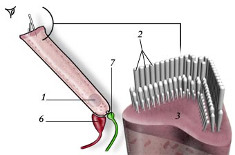 OHC OHC |
2. Stereocilia 3. Cuticular plate 4. Radial afferent ending (dendrite of type I neuron) 5. Lateral efferent ending 6. Medial efferent ending 7. Spiral afferent ending (dendrite of type II neuron) |
|
| In the human cochlea, there are 3,500 IHCs and about 12,000 OHCs. This number is ridiculously low, when compared to the millions of photo-receptors in the retina or chemo-receptors in the nose! In addition, hair cells share with neurons an inability to proliferate they are differentiated - this means that the final number of hair cells is reached very early in development (around 10 weeks of fetal gestation); from this stage on our cochlea can only lose hair cells. |

|
For
permission to non-commercial use of any element of this site, please contact us All rights reserved © 1999 - 2007 The authors Intellectual property law 85-660 (07/03/1985) |
|