| |
|
| Overview / Surface view / Stereocilia and Mechano-transduction | |
| Pictures : M. Lenoir | |
| Scanning electron microscopy (SEM) allows a clear surface view of the hair cells and of their stereocilia tufts. |
| |
|
| Overview / Surface view / Stereocilia and Mechano-transduction | |
| Pictures : M. Lenoir | |
| Scanning electron microscopy (SEM) allows a clear surface view of the hair cells and of their stereocilia tufts. |
 M. Lenoir |
 M.Lenoir |
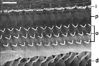
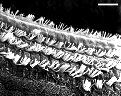
| The apical surface of inner support cells,
one row of inner hair cells (i: IHCs), pillars of Corti (p), 3 rows of outer hair cells (o: OHCs) and Deiters' cells are visible. At the bottom the surface is fractured and the base of the 3rd row of OHCs is seen, as well as phalangeal processes of Deiters' cell (d). See an electronic zoom scale bar: 15 µm |
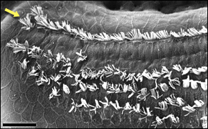
The surface of the organ of Corti is seen
at higher magnification and from the inner side so as to focus on the
stereocilia pattern of both IHCs and OHCs (see details on high magnification pictures below).
Also compare this very regular pattern with that observed at the apex.
scale bar: 10 µm |
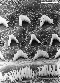
| Enlargements of IHC (left) and OHC (right) cuticular plates and stereocilia from rat basal turn | |
 M. Lenoir |
 M. Lenoir |
| Almost linear arrangement of IHC stereocilia | "W" pattern of OHC stereocilia |
| In both cases 3 rows of stereocilia of graded length, linked to each other, are embedded in a glabrous(i.e. bearing no microvilli) cuticular plate (in contrast with the surface of supporting cells which bear numerous microvilli). scale bar: 3 µm | |
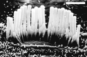
| SEM from the apical turn of a rat cochlea | |
 M. Lenoir |
In comparison with the basal turn, apical hair cells, especially OHCs, have stereocilia that are much longer, less regularly organised and much more loosely linked to each other. Frequently, as seen on the right, a 4th row of OHCs is observed. See an electronic zoom
|
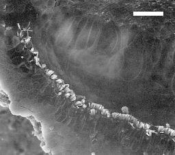
| Surface of apex of a guinea pig organ of Corti | |
 M. Lenoir |
IHCs (uppermost row) stay properly arranged in one single row up to the extreme apex, except for the last two cells (arrow). The distribution pattern of OHCs is greatly altered: 4 rows are seen in the right corner of the picture, but the organ of Corti ends (on the left) with 2, then 1 row of OHCs. scale bar: 15 µm. |
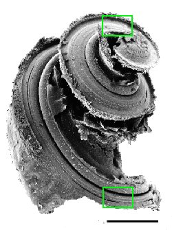
| Extreme apex of a mole rat cochlea | |
 M. Lenoi |
This apical disorganisation is even more pronounced in the mole-rat cochlea. Here, only the row of IHCs is seen, as though OHCs were not necessary for very low frequencies (below 20 Hz). scale bar: 15 µm |

|
For
permission to non-commercial use of any element of this site, please contact us All rights reserved © 1999 - 2007 The authors Intellectual property law 85-660 (07/03/1985) |
|