| |
|
| Overview / Synapses / Physiology | |
| Drawings: S. Blatrix; Pictures: R. Pujol | |
| |
|
| Overview / Synapses / Physiology | |
| Drawings: S. Blatrix; Pictures: R. Pujol | |
 R.Pujol R.Pujol |

This "synaptic
complex" is composed of: |
|
Typically, the synapse between IHC and auditory nerve endings is characterised by the presynaptic body surrounded by microvesicles (red arrow). Pre- and postsynaptic densifications of membranes are clearly visible (pink arrows), as well as an endocytotic profile (blue arrow) in the IHC. The glutamatergic nature of this synapse is discussed elsewhere. scale bar: 200 nm. |
|
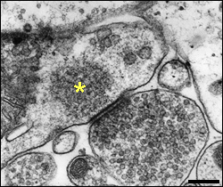
| The lateral efferent synapse | |
|
Vesicular endings of the lateral efferents synapse with afferent dendrites (from type I neurons connected to the IHC (see above). Note vesicle density in presynaptic ending and some dense material in the postsynapse (asterisk). scale bar: 100 nm |
 |
For
permission to non-commercial use of any element of this site, please contact us All rights reserved © 1999 - 2007 The authors Intellectual property law 85-660 (07/03/1985) |
|
 R.Pujol