| Overview / Spiral ganglion / Neurotransmitters | |
| Pictures : R. Pujol | |
| The spiral ganglion is formed from the primitive otocyst. It differentiates very early, before the organ of Corti. In man, it is composed of 30 to 35,000 bipolar neurons of two main types. Large and myelinated type I neurons (accounting for more than 90%) are connected to inner hair cells; small and unmyelinated type II neurons are connected to outer hair cells. Both types have central axons delivering messages to the cochlear nuclei. |
| The two types of spiral ganglion neurons | |
|
|
|
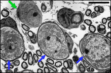
| Three type I neurons (blue
arrows) and one type II neuron (green arrow) are seen on the left, together
with sections of myelinated fibers (axons from neighboring neurons). On
the right, the bipolar nature of one type I neuron is better shown.
scale bars: 10 µm, left; 5 µm, right. |
|
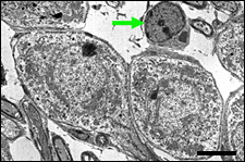
| Type I neuron and its satellite glial cell |
|
|
|
This type I neuron is ensheathed by processes
from a satellite glial cell which form a thin myelin sheath. Note the
dense cytoplasmic content, with numerous mitochondria, indicating the
high metabolic status of the cell. On the right is the axon hillock.
|
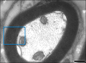
| Myelinated fiber |
|
|
|
Cross-section of a myelinated intra-ganglionic
auditory fibre (axon from a type I neuron). Within the axon, 3 mitochondria
and numerous microtubules are seen. The myelin sheath is formed of about
30 layers (see electronic
zoom). |
| Early stage of myelination |
|
|
|
During development, intra-ganglionic auditory
fibres (asterisks), are ensheathed by a glial cell process. Two of these
glial cells (yellow arrows) have already formed a myelin sheath of several
layers (see electronic zoom). |


|
For
permission to non-commercial use of any element of this site, please contact us All rights reserved © 1999 - 2007 The authors Intellectual property law 85-660 (07/03/1985) |
|