| |
|
| Overview / External ear / Middle ear / Inner ear | |
| Drawings: S. Blatrix | |
| |
|
| Overview / External ear / Middle ear / Inner ear | |
| Drawings: S. Blatrix | |
|
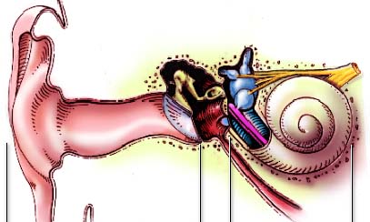
See higher magnification
drawings for details of the external,
middle and inner ear The animated drawing below illustrates the functional relationships of the 3 parts of the ear. |
||||
|
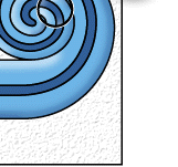
Here, on the right, a low frequency sound affects a more apical part of the cochlea |
|
 |
For
permission to non-commercial use of any element of this site, please contact us All rights reserved © 1999 - 2007 The authors Intellectual property law 85-660 (07/03/1985) |
|